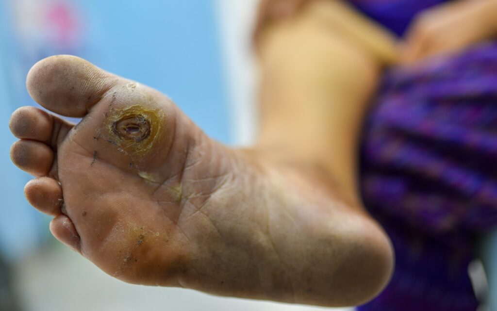
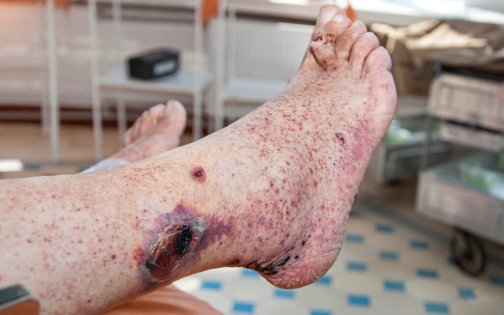
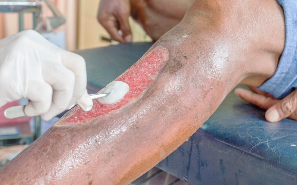
The treatment of chronic wounds is complex, time-consuming, and often costly. Therefore, it is essential to diagnose the underlying cause of the difficulty in healing a hard-to-heal wound, enabling causal treatment rather than just symptomatic action. This is particularly important, as it is believed that no venous or arterial ulcer will heal spontaneously without proper treatment. The same applies to wounds and ulcers associated with diabetic foot syndrome or other conditions.
The highly miniaturized device is capable of multimodal imaging of hard-to-heal wounds. It is important to emphasize that the wound scanning is performed in a contactless manner, ensuring the procedure is sterile, hygienic, and painless for the patient, while also allowing for a fast acquisition process.
The spectrum of imaging capabilities
of the scanner are:
• hard-to-heal wounds,
• post-operative wounds,
• diabetic foot,
• burns,
• ulcers,
• oncological lesions
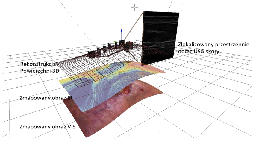
The broad scope of analysis determines the device's potential for application across various fields of medicine:
Rigid and soft surgery
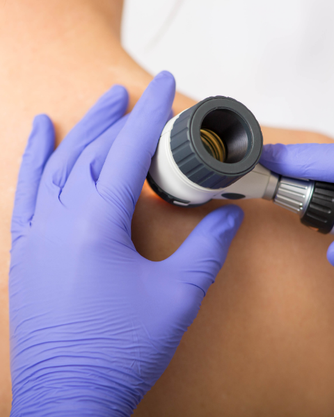
Dermatology

Oncology
March 19, 2024
April 21, 2023
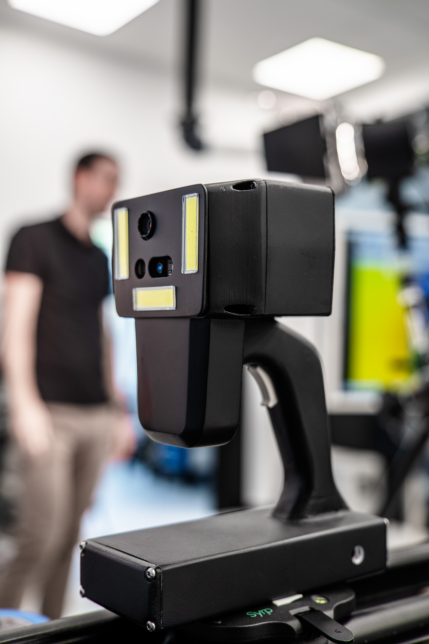
The assembly prototype of the wound scanner housing has been developed to facilitate the assembly and testing of the functionality of all electronic components. Following a thorough verification of the efficiency of individual digital modules, optimization of the device’s geometry is planned. The primary objective of this process is to reduce the overall size of the device while enhancing its aesthetics and user ergonomics.
April 28, 2023
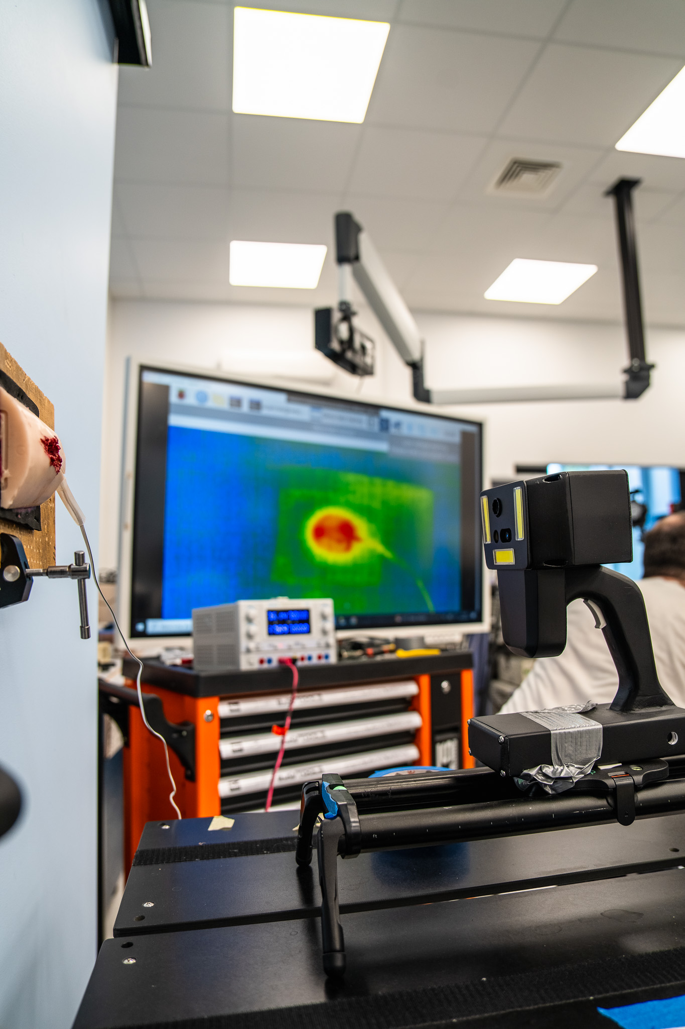
The WoundScanning device offers innovative wound scanning using thermography, enabling precise analysis and monitoring of treatment progress. With advanced technology, the scanner integrates various measurement spectrums into a single coherent image, significantly enhancing diagnostic accuracy and objectifying the treatment process.
Wound scanning with thermography provides numerous benefits, including:
– Infection Detection,
– Contactless Operation,
– Accuracy,
– Monitoring,
– Hydration Assessment,
– Speed,
– Painless Procedure,
– Cost-effectiveness.
September 15, 2023
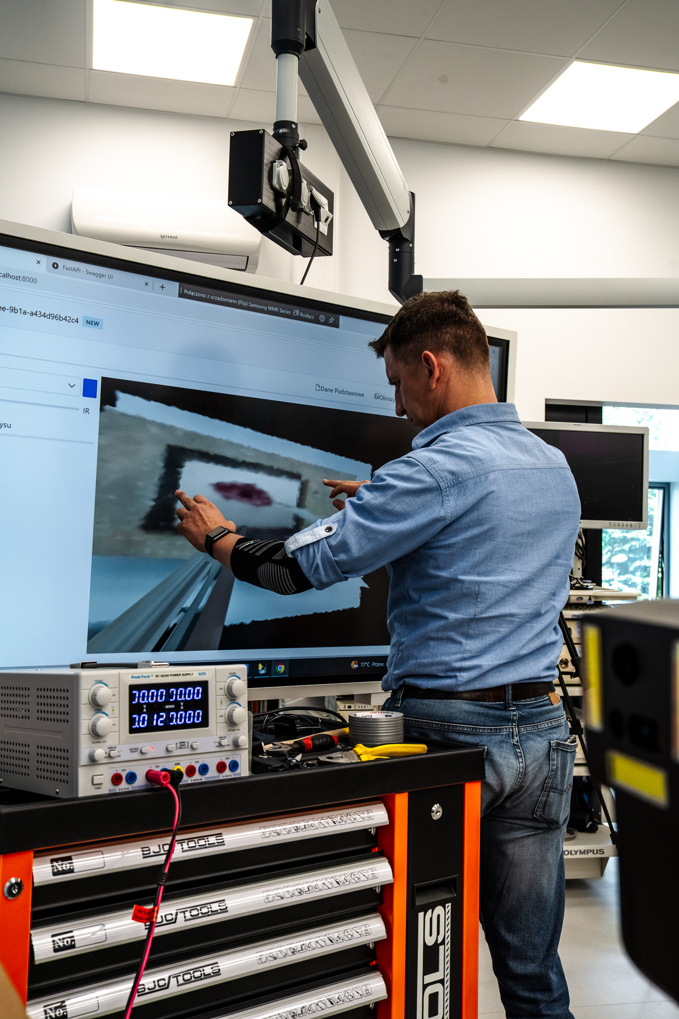
The developing wound scanner allows for 3D wound scanning using a ToF (Time-of-Flight) camera, providing an editable three-dimensional image. This modern technology integrates diverse measurement spectrums to create a coherent image, enabling more precise diagnosis and an objective treatment process.
3D wound scanning utilizing ToF technology offers numerous benefits, including:
– High Precision,
– Measurement Speed,
– Contactless Operation,
– 3D Visualization,
– Volume Quantification,
– Integration with Other Technologies,
– Painless Procedure,
– Usability in Hard-to-Reach Areas,
– Accuracy in Various Lighting Conditions.

Our team
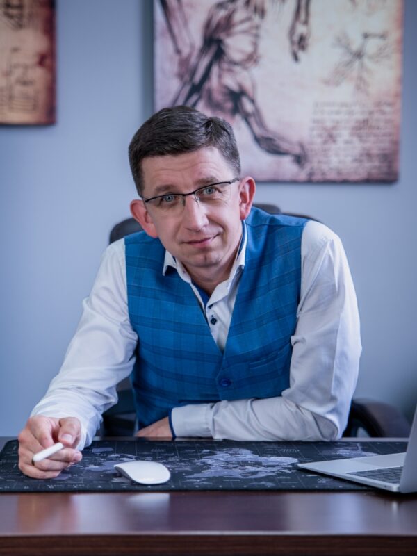
Marcin Sprawka
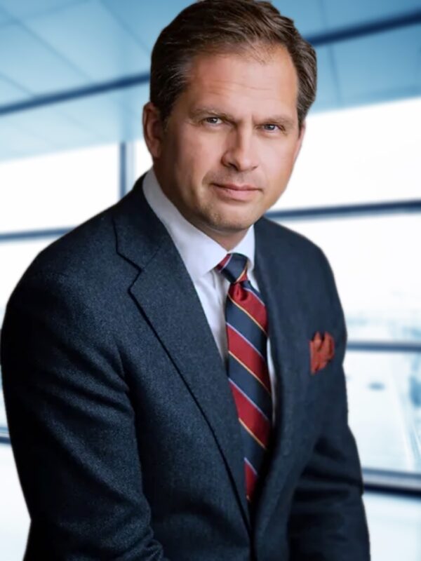
Prof. dr hab. n. med. Michał Zembala
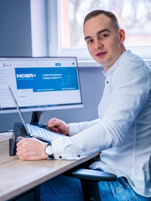
Mikołaj Dracz
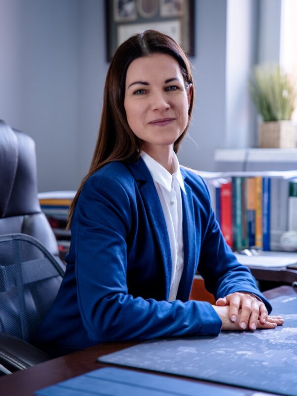
Katarzyna Ciesek
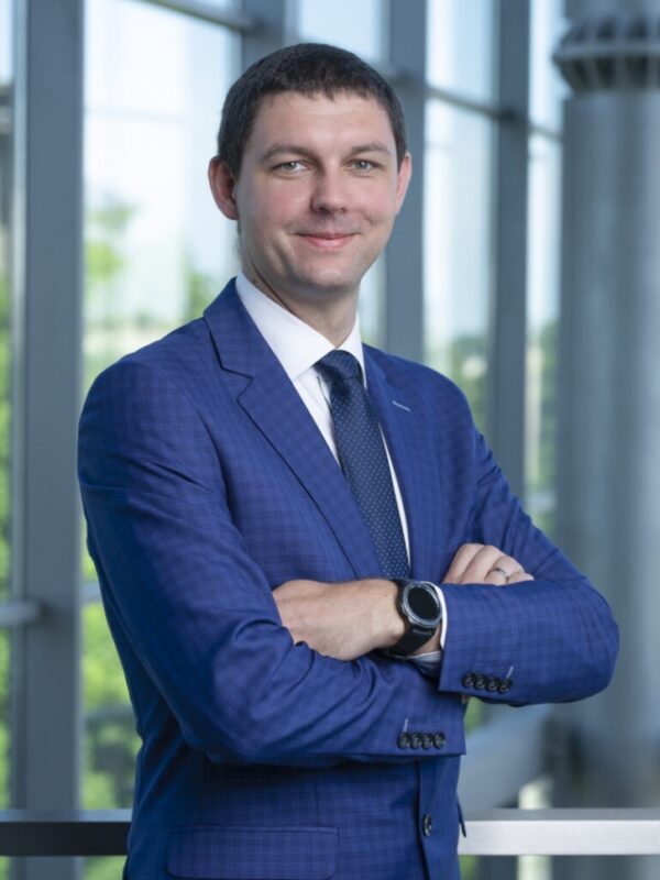
dr Jan Juszczyk
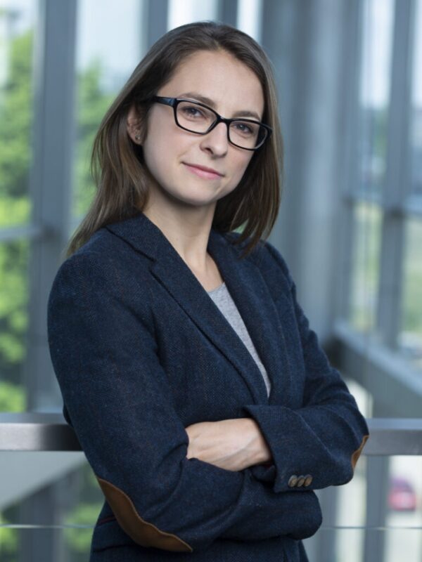
dr inż. Maria Bieńkowska
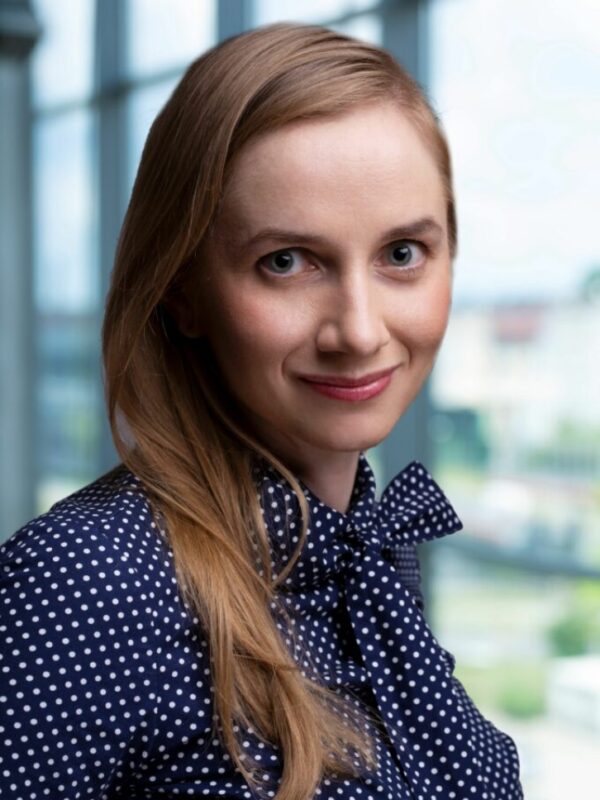
dr inż. Agata Wijata

mgr inż. Jacek Andrzejewski

WOUNDSCANNING
to opracowywane obecnie rozwiązanie służące do skanowania ran wraz z oprogramowaniem do realizowania fuzji obrazów termograficznych i fotograficznych oraz wykonywania obrysów 3D ran. Poprawa diagnostyki trudno gojących się ran oraz optymalizacja procesów leczenia jest priorytetem projektu.
URZĄDZENIE WOUNDSCANNING POZWOLI NA ANALIZĘ POLA RANY
W TRZECH SPEKTRACH:
Leczenie ran przewlekłych
jest złożone, czasochłonne
i często drogie.
W związku z tym należy zdiagnozować przyczynę trudności w wyleczenia trudno gojącej się rany, co pozwoli działać przyczynowo,
a nie podejmować działania wyłącznie objawowe. Zwłaszcza, że uważa się, że bez odpowiedniego leczenia żadne owrzodzenie żylne
lub tętnicze nie zagoi
się samoistnie. To samo dotyczy ran i owrzodzeń
w zespole stopy cukrzycowej czy innych.
Spektrum możliwości obrazowania skanera to:
• trudno gojące się rany,
• rany pooperacyjne,
• stopa cukrzycowa,
• odleżyny,
• oparzenia,
• owrzodzenia,
• zmiany onkologiczne
SZEROKI ZAKRES ANALIZY DETERMINUJE MOŻLIWOŚĆ ZASTOSOWANIA URZĄDZENIA NA WIELU OBSZAR MEDYCYNY:
ZESPÓŁ

Marcin Sprawka

Mikołaj Dracz

Prof. dr hab. Michał Zembala

Katarzyna Ciesek

dr Jan Juszczyk

dr inż. Maria Bieńkowska

dr inż. Agata Wijata


